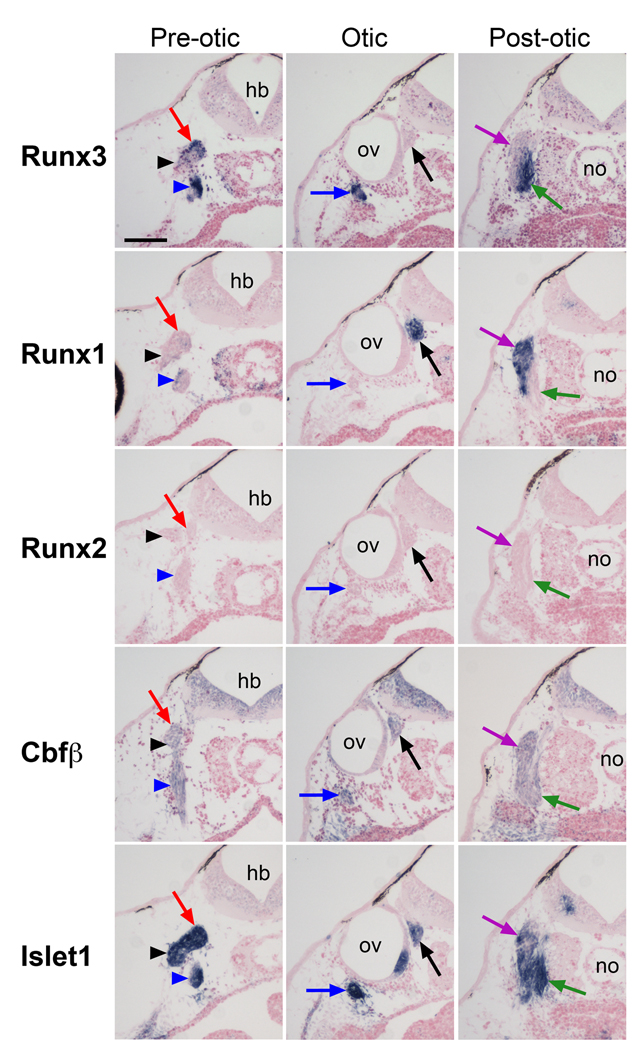
Figure 7. Comparative analysis of Runx3, Runx1, Runx2, Cbfβ and Islet1 expression in neurogenic placode derivatives at stage 40.
Transverse sections were performed at three levels along the antero-posterior axis; the pre-otic, the otic, and post-otic regions. Each column shows sections from approximately the same level. Trigeminal ganglion (red arrows), ganglion of anterodorsal lateral line nerve (black arrowheads), ganglia of facial and anteroventral lateral line nerves (blue arrowheads), ganglia of glossopharyngeal and middle lateral line nerves (blue arrows), statoaccoustic ganglion (black arrows), ganglion of vagal nerve (green arrows), ganglion of posterior lateral line nerve (purple arrows). hb, hindbrain; no, notochord; ov, otic vesicle; The scale bar in the upper left panel represents 100 µm.

