Fig. 7.
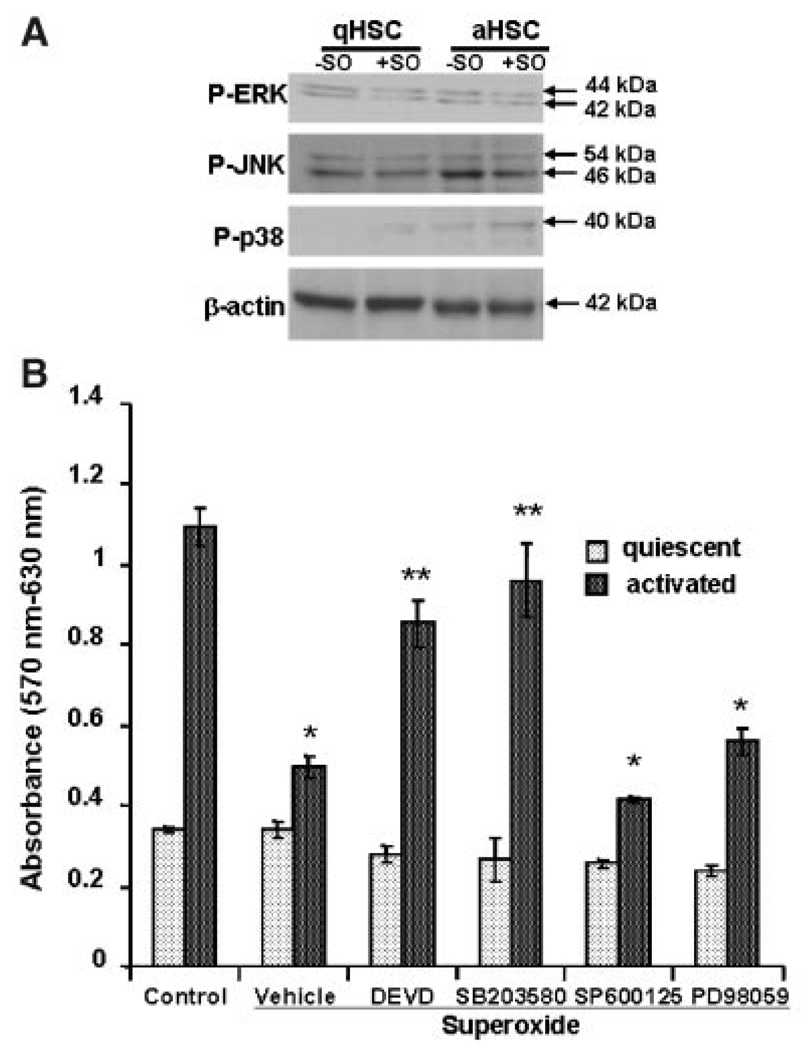
A: Effect of superoxide on activation of MAPKs. qHSCs or aHSCs were treated with superoxide for 3 h, and the lysates (20 µg protein) were subjected to SDS–PAGE followed by immunoblotting with antibodies against P-ERK, P-JNK, and P-p38. Equal loading was ensured by β-actin expression. B: Effect of pretreatment with caspase-3 and MAPK inhibitors on viability of HSCs incubated with superoxide. qHSCs or aHSCs were incubated with inhibitors of the activation of caspase-3 (DEVD-fmk), p38 (SB203580), JNK (SP600125), or ERK1/2 (PD98059) (all at 10 µM concentration) for 30 min prior to the addition of 1 mM HX ± 2 mU/ml XO. MTT assay was performed at 24 h. *P<0.01 versus control; **P<0.05 versus control and superoxide.

