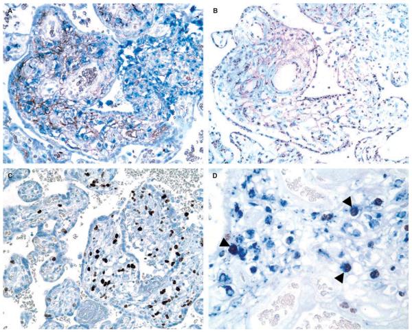
Figure 1.
A, Double staining for CD14 (brown) and CD68 (blue) detects an increased number of macrophages in the villitis of unknown etiology (VUE)-affected area. B, Chromogenic in-situ hybridization using a Y chromosome-specific probe (DYZ1) shows that a majority of the macrophages have a distinct intranuclear hybridization signal indicating that they are of fetal origin, whereas intervillous mononuclear inflammatory infiltrates of maternal origin are consistently negative (right upper part). C, VUE-affected areas are easily recognized by prominent Ki67 immunoreactivity in the inflammatory cells of the villous stroma (right side), when compared with unaffected areas (left side). D, Double immunohistochemistry for Ki67 (brown) and CD68 (blue) shows proliferating macrophages with Ki67 nuclear labeling (arrowheads).

