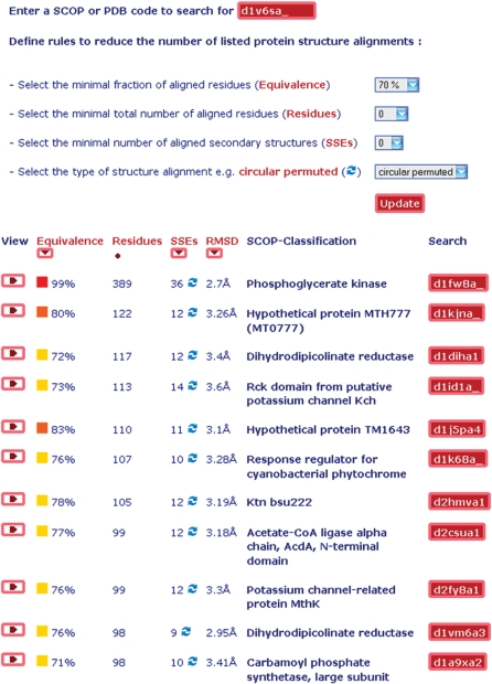
Figure 4.
Structure alignment browser (all-against-all) available at http://agknapp.chemie.fu-berlin.de/gplus. Top part: rules to search and reduce the number of listed protein structure alignments. Bottom part: sorted list of selected protein structure alignments with >70% of aligned residues <4 Å RMSD for the phosophoglycerate kinase with PDB id 1V6S, chain A relative to all proteins of the ASTRAL40 (SCOP 1.75) database of 10 444 protein structures.

