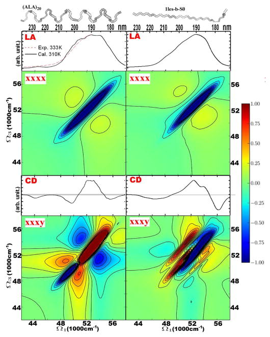
Figure 1.
Dependence of the linear and 2D signals on the backbone configuration: helical (left column) versus extended strand (right column). From top to bottom: structures, absorption, 2DUV xxxx, CD, 2DUV xxxy. The extended strand is taken from lectil lectin (Thr1-Phe11 of chain C in 1les.pdb, from Protein Data Bank) and the α-helical conformation is a 20 residue poly-alanine chain. The 2DUV spectra normalization (R) factors: for helical xxxx – 0.35, xxxy – 0.16; strand xxxx – 0.38, xxxy – 0.31.

