
Fig. 2.
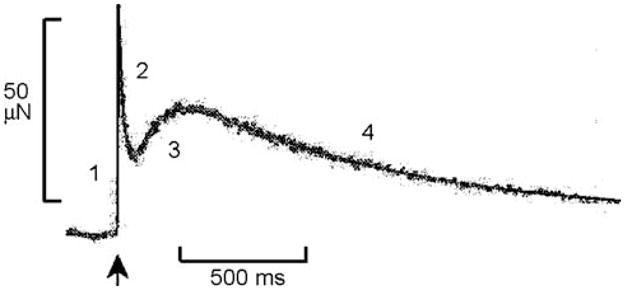
A record of the force time course that occurs in response to a step-length increase (at ↑, 1.5 nm/half sarcomere, which is about 0.12%) in rabbit psoas fibers at 5°C during Ca2+ activation. Numbers indicate four phases of tension transients. Modified from Fig. 3 of Davis et al. (2002), and reproduced with permission from the Biophysical Society