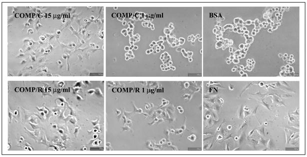
FIGURE 2. Morphology of TC1a attached to COMP/TSP5.
TC1a cells were allowed to attach to COMP/TSP5 coated at the indicated concentrations in the presence of 1 mM CaCl2 (COMP/C) or 5 mM EDTA followed by reduction (COMP/R). FN coated at 10 μg/ml (FN) or heat-inactivated bovine serum albumin (BSA) coated at the same concentration was used as control. Attachment assays were carried out as described under ”Materials and Methods.“ At the end of the 2-h incubation, phase-contrast pictures were taken.

