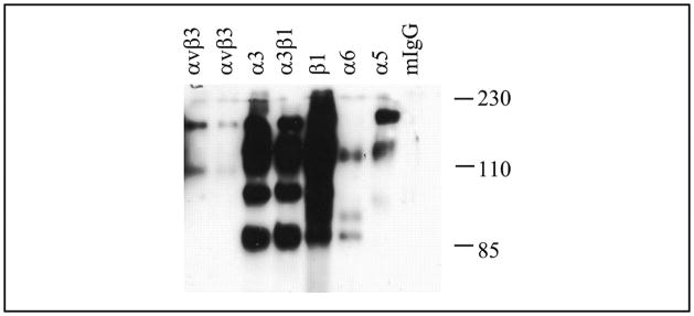
FIGURE 5. Integrin expression by the chondrocytes.
Primary chondrocyte cell surface proteins were labeled with biotin. The cells were lysed, and integrins were immunoprecipitated using specific integrin antibodies as indicated. The immunoprecipitated integrins were separated by SDS-PAGE followed by transfer to a piece of nitrocellulose membrane. The biotin-labeled integrins were visualized by horseradish peroxidase-conjugated streptavidin incubation followed by enhanced chemiluminescence detection. mIgG, mouse IgG.

