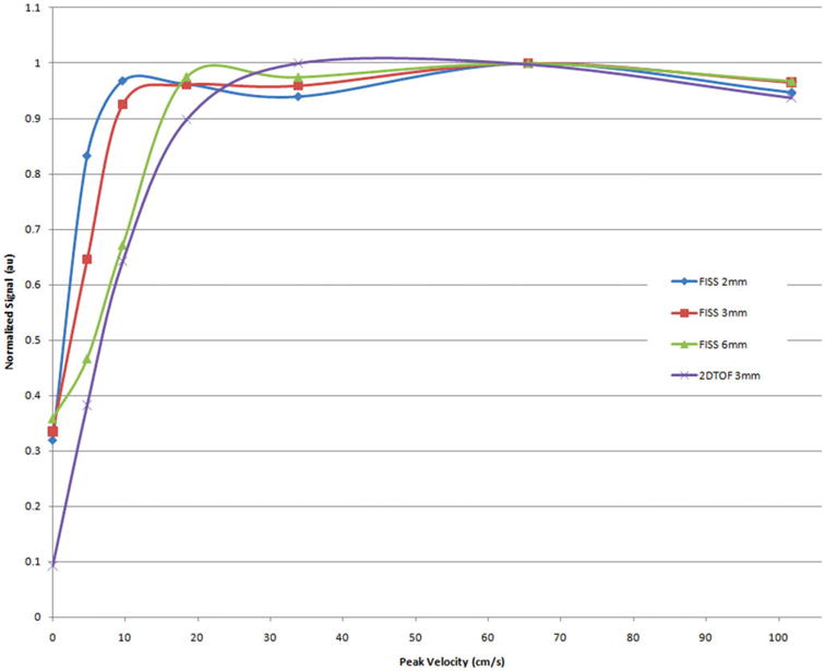
FIG. 7.

Relationship of signal to flow velocity for QISS (slice thicknesses of 2mm to 6mm) and 2DTOF (slice thickness 3mm) in a pulsatile flow phantom. The flow phantom had a triphasic waveform mimicking the pattern of normal peripheral arterial blood flow with a simulated heart rate of 71 beats/min. Note that the flow signal is approximately constant for QISS over nearly the full range of tested velocities, whereas the flow signal for 2DTOF drops precipitously for velocities less than 30 cm/sec. This helps to explain the observation in Fig. 6 that small branches and distal vessels, which have lower flow velocities than the larger proximal arteries, are not well shown with 2DTOF.
