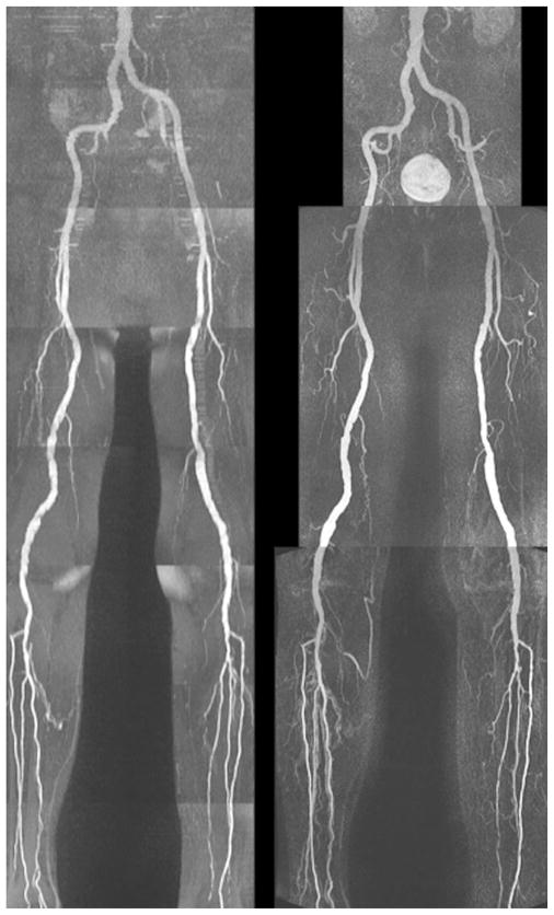
FIG. 8.
QISS MRA (left) versus CE-MRA (right) in a patient with vessel ectasia and mild multifocal disease. There is good correspondence between the two imaging tests. On the full-thickness MIP of the QISS MRA, a small focus of signal decrease was observed in the proximal left superficial femoral artery but appeared normal on thin-section MIPs (not shown).

