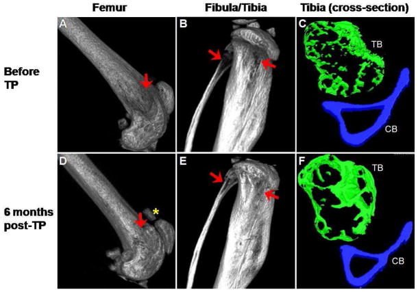
Figure 4.
Micro-CT images of femur, fibula and tibia from osteogenesis imperfecta murine (oim) mouse before and 6 months after transplantation of purified HSCs. The irregular bone structure in the femur before transplantation (arrow, A) improved after transplantation (arrow, D). Calcification of the tendon attaching to the patella was again noted after transplantation (asterisk, D). Similarly, the gaps in fibula and tibia (arrows, B) were filled after transplantation (arrows, E). Comparison of the cross-sectional images of tibia before (C) and after (F) transplantation highlights the extensive improvement in both trabecular (TB; green) and cortical (CB; blue) bone. The images in A, B, D and E are shown at ~ 100% magnification, C and F are shown at ~ 290% magnification. Correlative images are all displayed using the same threshold values.

