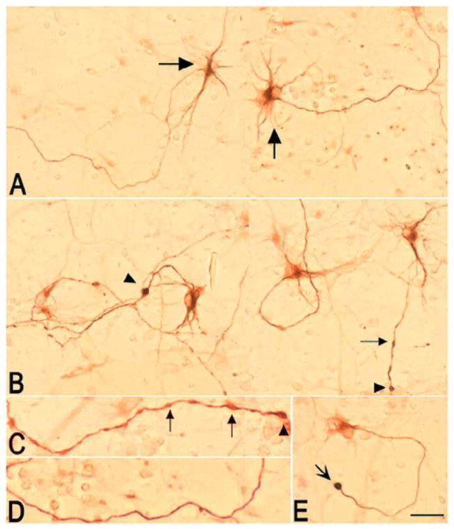
Figure 2. Effects of CE on the morphology of SMI-32 immunoreactive neurites in spinal cord neuronal cultures.
A) Composite picture showing two fields of a control culture maintained in medium/vehicle. A subpopulation of cells with motor neuron-like morphology (big arrows) and their processes are SMI-32 positive as reported previously (42). B) Composite picture showing two fields of a culture treated with 5 μM CE for 4 h. Addition of CE, induced beading (small arrows) and swelling (arrowhead) of neurites. C) High magnification picture of a neurite in a CE-treated culture showing beadings (small arrows) and swellings (arrowheads). D) High magnification picture of a neurite in a control culture showing a smooth process. E) An SMI-32 positive cell in a CE-treated culture showing a spherical structure resembling a retraction bulb (concave arrow). The experiment was repeated twice and yielded similar results (n=8). Bar = 100 μm.

