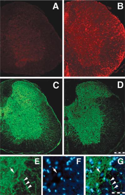
Fig. 8.
Mac-1 (A and B) and PMCA2 (C, D and E) immunostaining in lumbar spinal cord coronal sections in controls (A, C and E) and EAE (B and D) mice. Expression of Mac-1, a marker of microglia/macrophages, is increased throughout the whole lumbar spinal cord in EAE animals (day 24 PI) while the intensity of PMCA2 labelling is reduced. PMCA2 immunostaining is mainly localized to grey matter, with some labelled processes penetrating the ventral and lateral funiculi. (E–G) High magnification picture of PMCA2 (E), DAPI staining (F) and merged stainings (G) at the grey matter/white matter interface showing PMCA2 immunostaining on neuronal membrane (E, G, arrow) and some processes penetrating the white matter (E, G, arrowhead). Arrow in F indicates a typical neuronal nucleus stained with DAPI. Scale bar, 200 μm (A–D); 50 μm (E–G).

