
Figure 4.
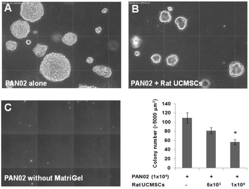
The colony growth of PAN02 pancreatic carcinoma cells was significantly attenuated by co-culture with rat UCMSCs in soft agar. PAN02 cells (1 × 10⁴) were mixed with either 5 × 10³ or 1 × 10⁴ rat USMSCs in the 0.45% top agar layer containing 0.04% Matrigel and 10% FBS and seeded on top of the 0.9% bottom agar layer. Colony growth was evaluated 10 days after seeding the cells. The pictures show the morphology of the PAN02 colonies in the presence (A) or absence (C) of 0.02% Matrigel or PAN02 colonies co-cultured with rat UCMSCs in the presence of 0.02% Matrigel (B) in soft agar after 10 days. The bar graph shows the summarized results from the colony assay carried out in the presence of 0.02% Matrigel. The experiment was performed twice with triplicate determinations. Although the total number of PAN02 and rat USMSCs combined was larger than in the control, the colony size and number were significantly smaller than in the control. The error bars represent the standard error of the mean of the samples. *, p≤0.05, as compared to the level of control.