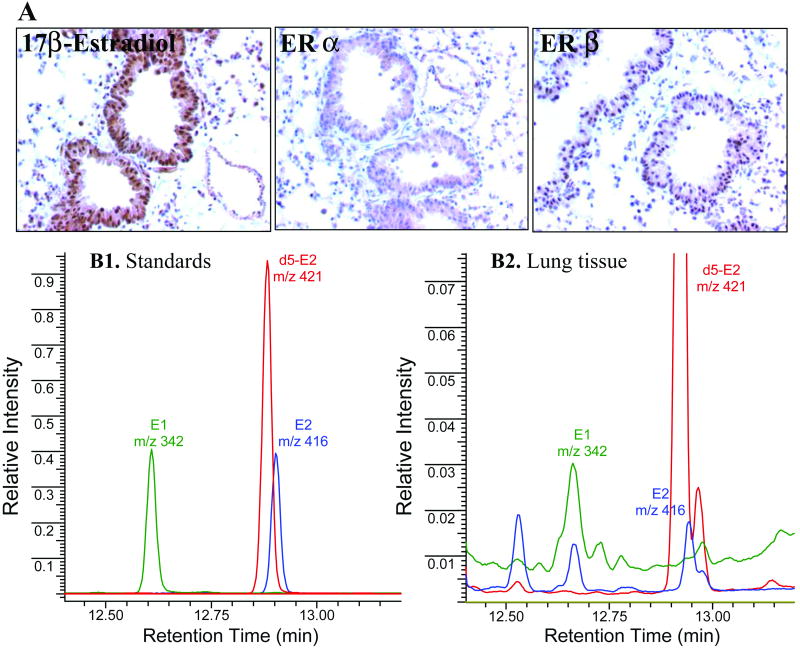
Fig. 4. Detection of estrogens within murine lung tissue.
Lung tissue from female A/J mice was subjected to immunohistochemical and GC/MS analyses. A. Detection of 17β-estradiol (E2), Erα and Erβ in lung epithelial cells by immunostaining. The bronchioloalveolar epithelium (BAE) stained positive for all antigens evaluated. Subcellular staining was observed as follows: E2, strong nuclear and cytoplasmic staining in the BAE and some pneumocytes; ERα, cytoplasmic staining of the BAE; and ERβ, nuclear staining in the BAE and some pneumocytes. B. Selective ion monitoring of trimethylsilyl derivatives of E1 and E2 (1.1 pmol each) and d5-E2 (2.6 pmol) as standards (B1) and in the murine lung tissue (B2). Each trace represents different ions monitored. Deuterium-labeled E2 represents the internal standard. Unmarked peaks in B2 denote unknowns; upper part of the chromatogram was cropped to enhance the visualization of small peaks.

