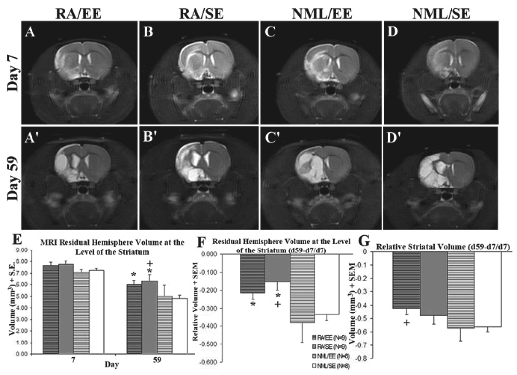
Figure 1. RA preserved hemisphere volume after stroke.
All animals had T2-weighted MRIs on day 7 (A–D) and day 59 (A'–D') after tMCAO and were assigned to treatment groups based on day 7 stroke volume (≥65% normal signal volume vs. intact hemisphere). All groups had similar stroke volumes prior to treatment (E). Rats that received an RA-enriched diet had more residual hemisphere volume at day 59 after MCAO compared with those on a normal diet (E, *p<0.05 vs NML/EE, +p<0.05 vs. NML/SE) as well as a smaller change in hemisphere volume over time (F, *p<0.05 vs NML/EE, +p<0.05 vs. NML/SE). Rats that received an RA-enriched diet and environmental enrichment maintained more striatal volume over time compared with normal controls (G, +p<0.05). All values are expressed as volume (mm3) ± SEM.

