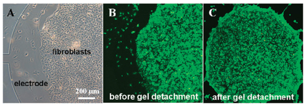
Fig. 4.
Cultivation of cells and release of cell-carrying hydrogels from ITO electrodes. (A) A bright field microscopy image showing fibroblasts adherent on hydrogel elements. The edge of the electrode as well as the lead (connection) are visible on the left side of the image. (B–C) Viability staining of cells adherent on heparin hydrogels before (B) and after (C) electrochemical detachment shows that cells are viable after detachment.

