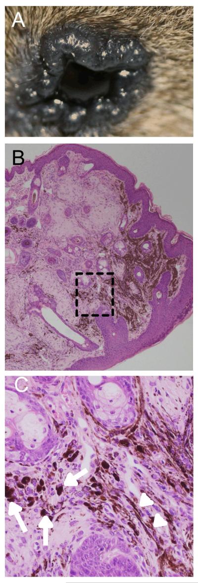
Figure 2. G12VKRAS induces pigmented epithelioid melanocytoma in mice.

A. Photograph showing an elevated dark lesion in the peri-orbital area of a TM-treated G12VKRAS;Tyr::CreERT2 mouse
B. Low power magnification of an H&E-stained paraffin section of a pigmented epithelioid melanocytoma from the peri-orbital area of a TM-treated G12VKRAS;Tyr::CreERT2 mouse. Note the absence of a junctional component.
C. High power magnification photomicrograph of the boxed area in B. Note the presence of heavily melanin-laden epithelioid cells (arrows) and less pigmented spindle cells (arrow heads).
