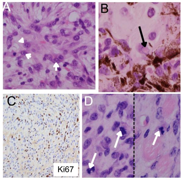
Figure 4. Characterisation of tumors induced by G12VKRAS.
A. A deep area from a perianal melanocytic lesion predominantly composed of hypo/amelanotic spindle cells exhibiting conspicuous nuclear pleomorphism and large nucleoli (arrowheads).
B. A deep area from a perianal melanocytic lesions composed of a mixture of spindle and epithelioid cells displaying the presence of pigmented cells. The arrow highlights an example of a nuclear pseudo-inclusion.
C. Ki67 positive staining within the deeper aspects of the neoplastic lesion.
D. Photomicrographs of mitotic figures (arrows) found within the deeper aspects of a perianal lesion.

