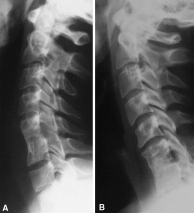
Fig. 4.

a Radiographic aspect at 12-year follow-up of a 61-year-old man treated with anterior decompression and fusion of the C5–C6 level, showing complete osteointegration of the bone graft with restoration of the physiologic lordosis and without radiographic evidence of degenerative changes at the levels adjacent. b Radiographic aspect at 13-year follow-up of a 59-year-old man treated with anterior decompression and fusion of the C6–C7 level. Early degenerative changes are noticeable at the C5–C6 level
