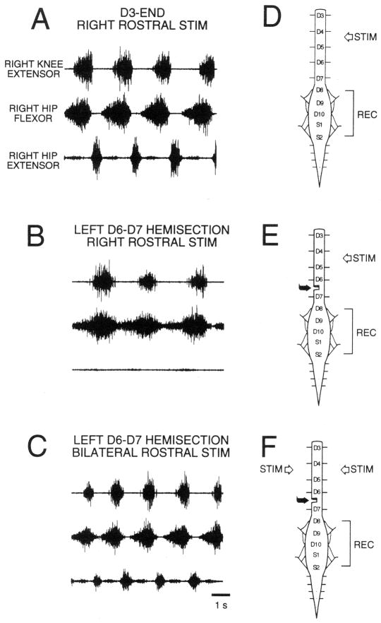
Figure 1.
Rostral scratch motor neuron activity patterns in the spinal immobilized turtle. Electroneurographic (ENG) recordings from the right biarticular knee-extensor nerve (top trace), the right hip-flexor nerve (middle trace), and the right hip-extensor nerve (bottom trace). (A) Right rostral scratch stimulation (STIM) in the D3-end preparation elicits normal rostral scratch with rhythmic right hip-flexor and hip-extensor alternation. (B) Right rostral scratch stimulation in the D3-end with left D6–D7 hemisection preparation elicits hip-extensor deletion rostral scratch with right hip-flexor rhythms. (C) Bilateral stimulation of mirror-image rostral scratch sites in the D3-end with left D6–D7 hemisection preparation elicits reconstructed normal rostral scratch with rhythmic right hip-flexor and hip-extensor alternation. (D) Sketch of the spinal cord in the D3-end preparation with a complete transection between the second and third postcervical spinal segments (D2–D3). (E and F) Sketches of the spinal cord in the D3-end with left D6–D7 hemisection preparation. All ENG recordings (REC) are from nerves on the right side. From48 and used with permission of and copyright 1998 by the New York Academy of Sciences and Wiley-Blackwell.

