Figure 5.
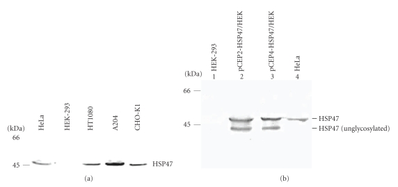
(a) HSP47 expression in different cell lines. Western blot analysis of conditioned cell lysates from different cell lines probed with monoclonal antibody to HSP47. Left, molecular mass standards expressed in kDa. (b) Western blot analysis of HSP47 expression in untransfected HEK-293 cells (lane 1), HEK-293 cells transfected with pCEP2-HSP47 (lane 2) or with pCEP4-HSP47 (lane 3) constructs and in HeLa cell lysate as positive control (lane 4). Two bands were detected in transfected HEK-293 cells lysates corresponding, respectively, to the glycosylated and unglycosylated forms. Left, molecular mass standards expressed in kDa.

