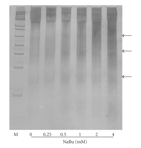
Figure 2.
Electrophoretic patterns of DNA isolated from NaBu-treated and untreated CHO cells. Isolated chromosomal DNA (2 μg) was electrophoresed on 5% polyacrylamide gel and intact and fragmented DNA bands were revealed by silver staining. Arrowheads designate DNA ladder formation. M: molecular weight marker, 100-bp DNA ladder. DNAs were isolated from day 8 cultures exposed to different concentrations of Nabu.

