
Fig. 3. Maintenance of the cone outer segment by RdCVF.
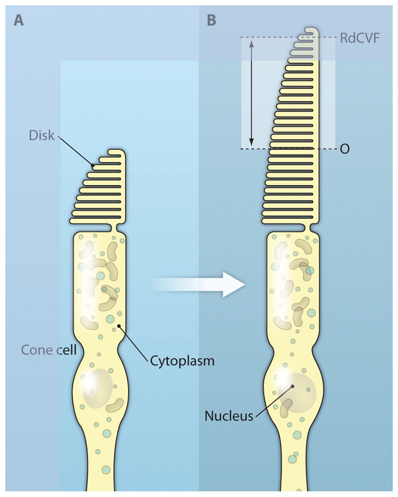
RdCVF was injected into eyes of the P23H rat, a model of autosomal dominant RP. The cone cells (green) of the RdCVF-injected animal (bottom panel) display a longer outer segment (indicated by double arrows) and smaller diameter (asterisk) than those in the vehicle-injected rat (top panel). The relative positions of the cone outer segment in each case are indicated on the diagram on the right.