
Figure 3.
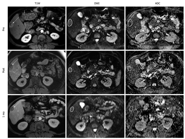
Representative T1W, DW, and ADC MR images from a 70 year-old man with right-lobe HCC secondary to autoimmune hepatitis. Images were acquired before, immediately after, and 1 mo after TACE. Arrows on the anatomical images indicate tumor location with sample ROIs drawn on the functional MR images. Intra-procedural ADC changed by 28.6%, which predicted a favorable EASL response at 1 mo. T1W: T1-weighted; DW: Diffusion-weighted.