
Figure 2.
Phenotype of the XLCOD5 Family
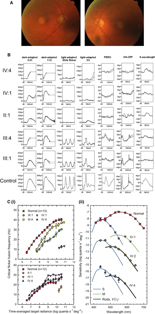
(A) Color fundus photographs of affected male patient II:3 (80 yrs old), showing symmetrical bilateral macular atrophy.
(B) PERG and full-field ERGs for affected males IV:4, IV:1, and II:1 and obligate carrier females III:1 and III:4, as indicated, compared with those from a representative normal subject (bottom row). Recordings were performed with gold-foil corneal electrodes. Broken lines replace eye-movement artifacts.
(C) L-cone cff measurements (i, upper panel) for two affected males (IV:1 and IV:4) and two obligate carriers (III:1 and III:4), showing significant loss of L- and M-cone sensitivity in affected males and reduced L-cone sensitivity in both carriers as compared to controls. S-cone critical flicker fusion measurements (i, lower panel) for three affected males (IV:1, IV:4, and II:1), showing significant S-cone sensitivity loss in the eldest affected male (II:1) as compared to controls. Spectral-sensitivity measurements (ii) for the three youngest affected males (IV:1, IV:2, and IV:4), showing substantial losses of flicker sensitivity at middle and long wavelengths, but sensitivities at short wavelengths that are nearly normal.
Error bars represent ±1 SEM.