
Figure 4.
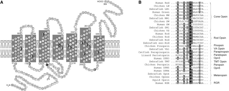
Secondary Structure of the Cone Opsins and Conservation of W177
(A) The secondary structure of the red (LW) and green (MW) cone opsins. Amino acid differences between the LW and MW opsins are shown as half-closed circles. W177 in transmembrane domain 4 and C203 are shown as a closed circle.
(B) Sequence alignment encompassing W177 of LW and MW opsins with known visual and nonvisual opsins in a variety of species, as named on the left. This tryptophan is 100% conserved in all opsins.