
Figure 4.
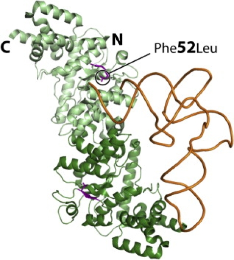
Interaction Model of Human YARS2 with tRNA
The model shows the crystal structure of the enzyme17 (without the S4-like C-terminal domain) on which one tRNATyr molecule has been docked according to its location in the crystal structure of a bacterial tryosyl-tRNA synthetase and tRNATyr complex.31 The two monomers are distinguished by dark- and light-green color with the first β strand encompassing residue 52 (p.F52L), shown as the mutant L52, highlighted in purple. Note the proximity of L52 with the accepting end of tRNA shown as orange.