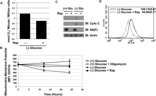
Figure 4. Mitochondrial activity is not increased in rapamycin treated TSC2-/- p53-/- MEFs.
A. TSC2-/- p53-/- MEFs were deprived of glucose for 24 hours, and the rate of oxygen consumption was measured. Oxygen consumption was measured by Clark's electrode, and the rate of consumption was determined and indicated as nmol of oxygen per minute per million cells. The experiment was conducted 3 times and yielded similar results.
B. Mitochondrial membrane potential was measure at 12 and 24 hours post treatment conditions via DiOC6 MFI via FACs. Oligomycin was used as a positive control (5μg/mL) in cells grown in glucose containing media. (n = 3)
C. PGC-1α and cytochrome c protein levels were determined at 24 hours post-deprivation.
D. Mitotracker measurement in the absence of glucose, and with or without rapamycin.

