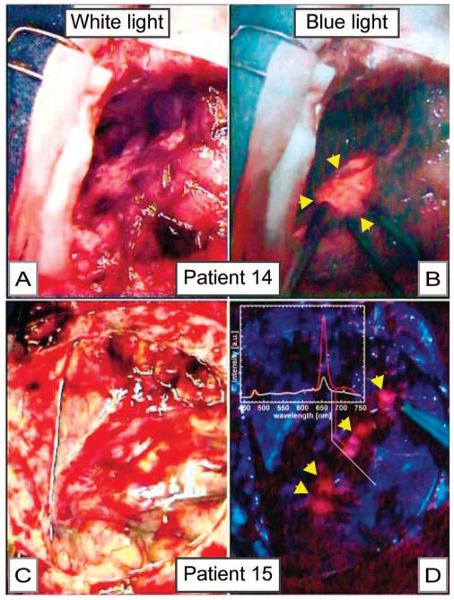
Figure 11.
Intraoperative images of a partially resected brain tumor (A and B) and the surface of the brain (C and D), comparing images obtained by white light (A and C) and mTHPC fluorescence imaging under blue light (B and D). In A, a partially resected tumor is difficult to discern while it is readily apparent in the blue-light mTHPC fluorescence image of the same tissue. Similarly, in C (a white light image of the surface of the brain), there is no apparent tumor, while the mTHPC fluorescence image, which is complemented by spectroscopy (inset) of the same field reveals the presence of malignancy. (Figure reproduced with permission from ref 80. Copyright 2001 American Society for Photobiology.)

