
Figure 12.
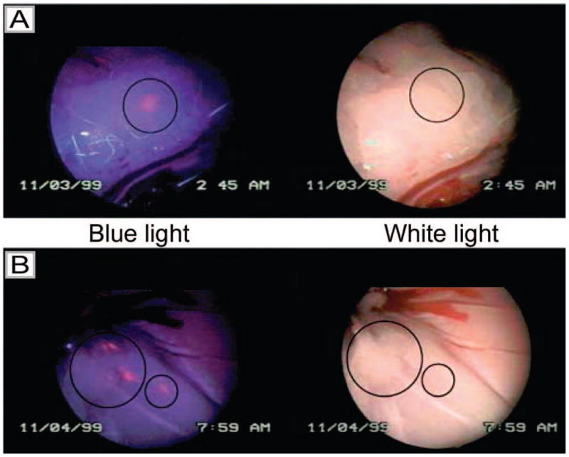
Laparoscopic images of peritoneal metastases in a rat model of ovarian cancer obtained by white light imaging (right) and HAL-induced PpIX fluorescence imaging under blue light illumination (left). Image (A) shows a lesion that is only visible in the blue light mode, but not by white light (position marked by a circle) (8mM HAL after 2 h). Image (B) shows three lesions visible by both blue and white light (big circle) and one only detectable by fluorescence (small circle) (8mM HAL after 2 h). (Figure reproduced with permission from ref 94. Copyright 2003 Cancer Research UK.)