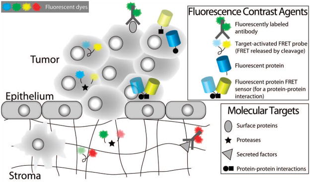
Figure 25.
Fluorescence contrast agents relevant for molecular imaging of biological responses to PDT. Fluorescently labeled antibodies, target-activated probes and genetically encoded fluorescent proteins are examples of fluorescence contrast agents that have been applied for molecular imaging of biological responses to PDT. This figure highlights the use of these contrast agents for detecting molecular factors in both the intracellular and extracellular spaces. The natural clearance of unbound antibody-fluorophore conjugates from the extracellular space enables their use for labeling cell surface proteins and extracellular secreted factors. Target-activated probes based on FRET (as shown here) or ground state quenching, are applicable for imaging both intracellular and extracellular factors. Fluorescent proteins (an endogenous labeling scheme) are useful for monitoring protein expression levels and protein-protein interactions, and for visualizing protein trafficking. Fluorescent protein FRET sensors also exist and can be used to detect protein-protein interactions. Examples of the application of these contrast agents for studying PDT-induced molecular mechanisms are shown in Figure 28 and are discussed in the text.

