
Figure 9.
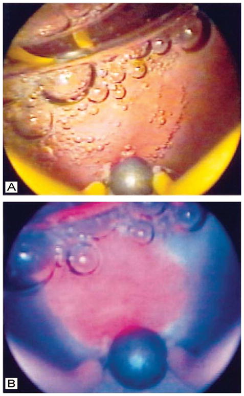
Comparison of white light and PpIX fluorescence images of an intermediate grade malignant lesion in the bladder of a human patient, obtained via cystoscopy. In the white light image (A), the lesion is not evident, while in the PpIX fluorescence image obtained under blue illumination (B), the lesion is readily visible as a pink region just above the large air bubble in the lower middle part of the field. (Figure reproduced with permission from ref 62. Copyright 1999 BJU International.)