
FIGURE 6. Lipid raft targeting of Cx43 and LC3 in cardiac tissue.
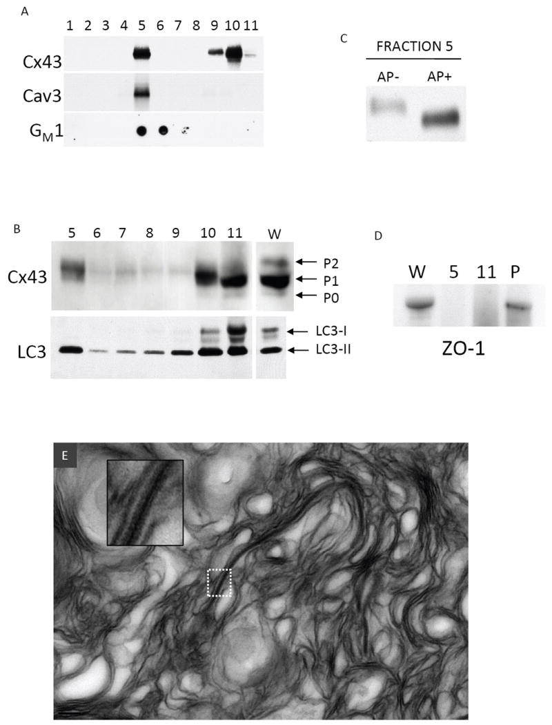
(A) Sucrose gradient fractionation of canine ventricular myocardium Western blotted for Cx43, caveolin-3 (Cav-3), and GM1 (dot blot probed with HRP conjugated cholera toxin B subunit). Fractions 1-4 correspond to 5% sucrose, 5-10 correspond to 38% sucrose, 11 corresponds to 40% sucrose. (B) Fractions 5-11 probed for Cx43 (separated by large format (18cm2) 10% acrylamide gel) and LC3. The lane marked W is a whole tissue lysate. Distinct phosphorylated species of Cx43 (P0, P1, P2) and LC3-I and LC3-II are indicated with arrows. (C) Cx43 Western blot of Fraction 5 that was untreated (AP-) or treated (AP+) with alkaline phosphatase and separated on a 10% acrylamide gel. (D) ZO-1 is absent from fraction 5 and 11, but was present in the pellet (P). (E) Material in fraction 5 was pelleted and prepared for TEM. A representative TEM of fraction 5 exhibits primarily multilamellar membranous material with an incorporated pentalaminar membrane (the inset is a higher power view of the boxed region).