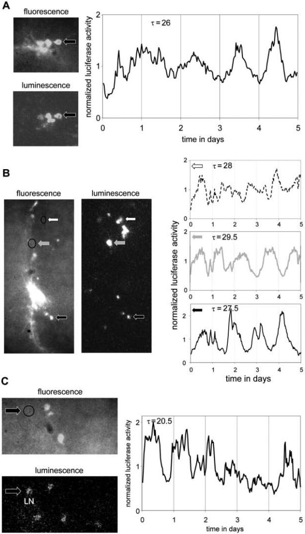
Figure 2.
Circadian imaging of individual clock neurons across different cell types and genotypes (see also Supplemental Material S3). Using the same fluorescent and bioluminescent reporters as in Figure 1 (Pdf>GFP and tim-luc) significant circadian luminescence rhythms were detected in individual ventral and dorsal lateral neurons (LNvs and LNds) in cultured brains from wild-type (A, B) or short period mutant clock mutant (dbts/+) flies (C). Period lengths (τ) of significant circadian luminescence rhythms during days 2 to 5 (p < 0.001; A, B) and 1 to 5 (p < 0.025; C) are indicated. Arrows and graphs are patterned in C to identify the cells whose luciferase rhythms are plotted. The absence of fluorescence in clock neurons imaged in B and C (circled areas) indicates that these are not PDF-expressing cells. Based on their location and morphology these cells were identified as LNd (B) or LN (either 5th sLNv or LNd; C).

