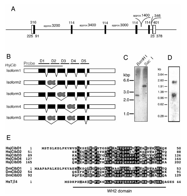
Figure 3.
Isoform structure of HsjCib gene. A: The gene structure of HsjCib inferred from PCR amplification of genomic DNA. Coding regions are represented in black. Numbers indicate the lengths (bp) of regions. B: Structure of obtained isoforms. Smaller isoforms were thought to skip some exons. The gray bar indicates the fragment used as probes in Southern, northern, and in situ hybridization. D1 to D5 indicates putative WH2 domains. C: Southern hybridization indicates that HsjCib exists as a single-copy gene on the genome. D: Northern hybridization for HsjCib. Several isoforms seem to overlap between the 0.96- and 1.38-kbp markers. The longer fragment around 2.5 kb may be a paralog or an isoform that has not been cloned. E: Alignment of putative amino acid sequences of HsjCib with Drosophila Cib and human Thymosin-β4.

