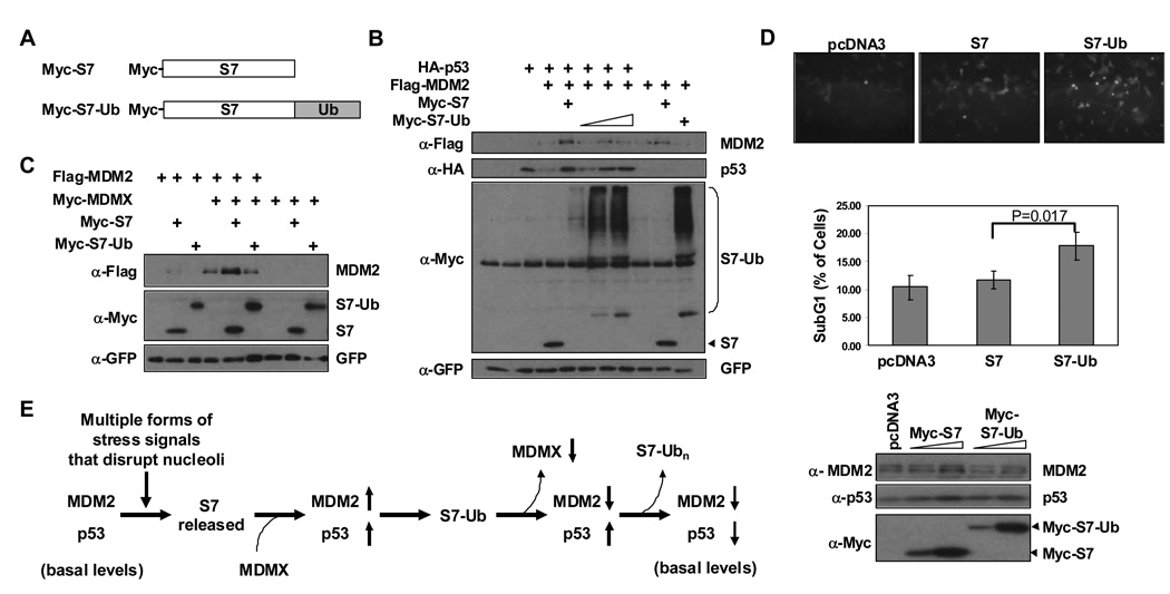
Figure 7. An S7-ubiquitin Fusion Protein Stabilizes p53 but not MDM2 and Promotes Apoptosis.
(A) Schematic representation of Myc-S7 and its derivative (Myc-S7-Ub) that has one copy of ubiquitin fused to the C-terminus. (B) S7-Ub stabilizes p53 more efficiently than MDM2. U2OS cells were transfected with the indicated combinations of HA-p53 (0.28 µg), Flag-MDM2 (1.6 µg), Myc-S7 (0.8 µg) or increasing amounts of Myc-S7-Ub (0.8, 1.6, 2.2 µg). Cell lysates were subjected to IB with indicated antibodies. GFP construct was added for transfection and loading control. (C) S7-Ub cannot cooperate with MDMX to stabilize MDM2. U2OS cells were transfected with the indicated combinations of Flag-MDM2 (0.6 µg), Myc-MDMX (0.6 µg), Myc-S7 (0.6 µg), or Myc-S7-Ub (2.2 µg). Cell lysates were subjected to IB with indicated antibodies. GFP construct was added for transfection and loading control. (D) S7-Ub induces more apoptosis than S7. U2OS cells were transfected with Myc-S7 (0.6 µg) or Myc-S7- Ub (2.2 µg) together with an F-GFP plasmid (0.1 µg). Thirty hours after transfection, cells were examined by immunofluorescence microscopy for floating cells (top panel) and then harvested for both FACS analysis (middle panel) and Western blot analysis (bottom panel). Apoptotic cell index and standard deviations were obtained from three independent experiments. P-value was calculated using Student’s t-Test with two-tailed distribution. Immunofluoresence and western blot images are representative data from these independent experiments. (E) A Model for S7 Regulation of the MDM2-p53 Circuit. Stress signals lead to ribosomal biogenesis perturbation and partial nucleolar disruption. S7 is released from nucleoli where it can interact with MDM2 leading to inhibition of MDM2-E3 ligase activity towards p53. MDM2 autoubiquitination is inhibited by S7 in an MDMX dependent manner. In addition, S7 serves as an E3 ligase substrate for MDM2 and since ubiquitinated S7 cannot stabilize MDM2, this may promote a feed-forward mechanism to maintain p53 mediated cellular response. Reduced levels of S7 would eventually allow p53 to return to basal levels.

