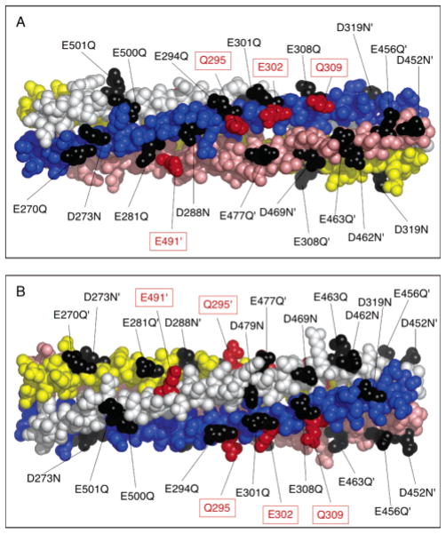
Figure 2.
Atomic model of the adaptation subdomain, highlighting the acidic side chains. The indicated structure is excerpted from the structural model for the full receptor (5). The helices of subunit I are colored yellow and pink, and the helices of subunit II are colored blue and white. Adaptation sites are colored red. The 17 acidic Asp and Glu side chains targeted in this study for neutralization by conservative mutation to Asn and Gln are colored black. (B) Alternate view obtained by rotating the subdomain 90° about its long axis. Structures were generated with MacPymol (DeLano Scientific).

