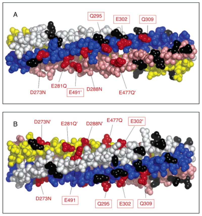
Figure 5.

Locations of the four newly identified activating sites, and of the four adaptation sites. The helices of subunit I are colored yellow and pink, and the helices of subunit II are colored blue and white. Adaptation sites are denoted in red and boxed. Positions where charge-neutralizing substitutions activate or have no effect on kinase activity are colored red or black, respectively. (B) Alternate view obtained by rotating the subdomain 90° about its long axis. Structures were generated with MacPymol (DeLano Scientific).
