Fig. 4.
Histological findings
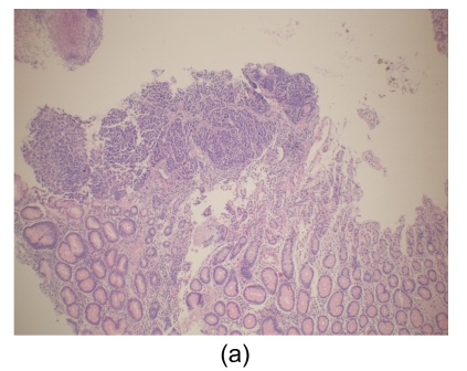
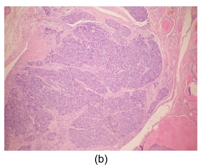
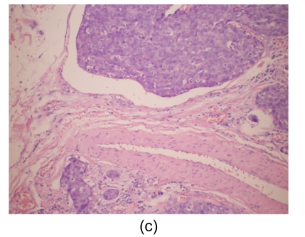
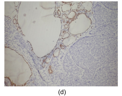
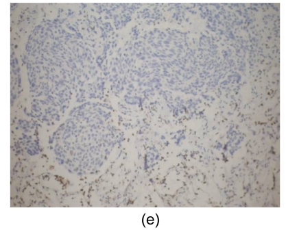
(a) Stomach. Tumor cells arise from the gastric mucosa (hematoxylin & eosin (H & E) staining, magnification 40×); (b) & (c) Thyroid. Multi-focal nests of tumor cells are distributed between the follicles (b) (H & E staining, 40×) and in the vascular lumen (c) (H & E staining, 200×); (d) & (e) Lung. Negative staining for thyroid transcription factor-1 (TTF-1) (d) and thyroglobulin (e) in tumor cells (200×)

