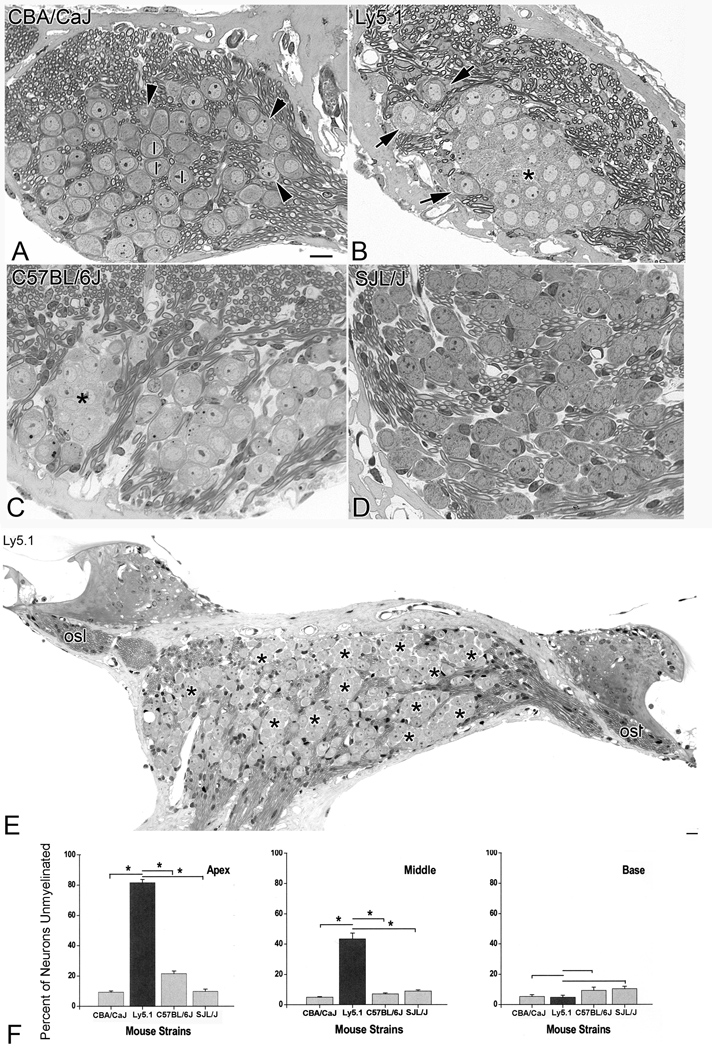
Figure 1. Morphological and quantitative evaluation of SGNs.
A. SGNs in the apical turn of a young adult CBA/CaJ mouse. The majority of neurons are myelinated type I (I) neurons, which can be distinguished from type II (arrowheads) neurons by their round nucleus and darker cytoplasm. The perikaryon of type I cells is surrounded by a dark line, which represents the myelin sheath. Type II neurons are of smaller size, have lighter cytoplasm and eccentric nuclei, and are located near the root of the osseous spiral lamina or close to the intra-ganglionic spiral bundle. Note that few neurons are in direct contact with each other. Most neurons are separated by extracellular space or nerve fibers. B. SGNs in the apical turn of an Ly5.1 mouse. The majority of neurons are unmyelinated and grouped together as a cluster (*). The aggregated neurons have round nuclei, dark cytoplasm and lack a myelin sheath. No myelinated nerve fibers are visible within the clusters. Three myelinated type I neurons (arrows) are located at the periphery of the neuronal cluster. C. SGNs in the apical turn of a C57BL/6J mouse. A small cluster of eight unmyelinated neurons (*) is located on the left side of the section. Somata of these unmyelinated cells are clumped as shown in Fig.1B. Note the numerous normal-appearing myelinated type I neurons scattered in other areas of Rosenthal’s canal. D. SGNs in the apical turn of a SJL/J mouse. The majority of neurons are myelinated type I neurons. No neural aggregation was seen in this strain. E. Unmyelinated and aggregated neurons in the apical (right side) and middle (left side) turns from an Ly5.1 mouse. The neural clusters are indicated with asterisks. The myelinated fibers in the osseous spiral lamina (osl) appear normal. F. Mean percentages of unmyelinated neurons in the apical, middle and basal turns of CBA/CaJ, Ly5.1, C57BL/6J and SJL/J mice. Unmyelinated cells comprise about 80% of SGNs in the apical turn and 40% of SGNs in the middle turn of Ly5.1 mice. Differences in the percentage of unmyelinated neurons between the Ly5.1 and the other three strains are statistically significant in the apical and middle turns (p< 0.01). Scale bars: A–D, 10 µm; E, 20 µm.

