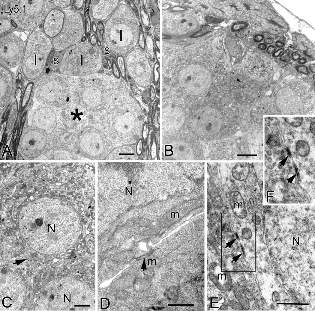
Figure 2. Ultrastructure of neural clusters in Ly5.1 mice.
A. Aggregated unmyelinated SGNs in the middle turn form a cluster (*) with a common myelin sheath. Neurons within the cluster have round nuclei and mitochondria-rich cytoplasm. Several normal-appearing myelinated type I SGNs (I) separated by Schwann cells (S) and neural processes occupy the region above the cluster. B. Neural cluster in the apical turn of another Ly5.1 mouse. Note that several nerve fibers appear outside the neural cluster. C. Aggregated unmyelinated neurons with large round nuclei (N). Their perikarya are directly in contact with one another. A black arrow points to a specialized membrane structure between neighboring cells. D. and E. Higher magnification images of somato-somatic apposition revealing junction-like symmetrical structures. The arrows point to the dark patches on the adjoining plasma membranes of two neighboring neurons. F. Enlarged image of corresponding boxed area in Fig. 2E. m, mitochondria. Scale bars: A and B, 2.5 µm; C, 1.5 µm; D and E, 500 nm.

