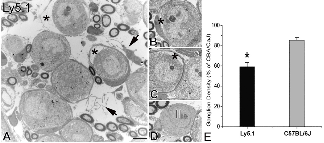
Figure 3. Neural loss in the basal turn of Ly5.1 mice.
A. Histopathological changes in the spiral ganglion. Two neurons have apparently separated from their surrounding myelin sheath (*). A homeless Schwann cell and Schwann cell processes appear within the extracellular space (arrows). B and C. Separation of Schwann cells from type I neurons (*). The neurons were in the basal turn taken from another Ly5.1 mouse. D. Type II neuron in the basal turn. The image was taken from the same mouse shown in Figs. 3B and C. Note that non-myelinating Schwann cells appear around the neuron. E. Mean ganglion cell density in the basal turn of Ly5.1 and C57BL/6J mice expressed as a percent of that in CBA/CaJ mice. The loss of SGNs in Ly5.1 mice is significant (p< 0.01). Scale bars: A–D, 2.5 µm.

