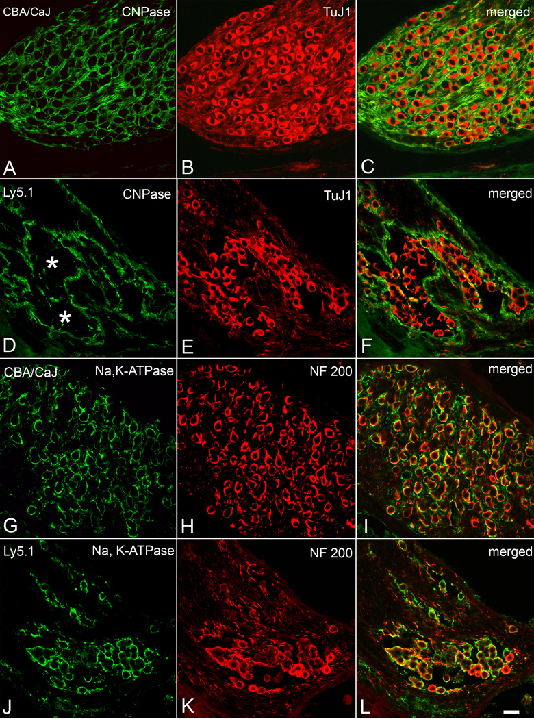
Figure 4. Immunostaining for neuronal and glial markers in apical turns from a CBA/CaJ and an Ly5.1 mouse.
A–C. Dual labeling with anti-CNPase (green) and anti-TuJ1 III β-tubulin (red) in a CBA/CaJ mouse. TuJ1 antibody preferentially labels the cytoplasm of large type I neurons (Sekerkova et al., 2008). CNPase labeling of Schwann cells and their myelin sheaths results in a honeycomb-like pattern in the SG. D–F. Dual labeling with anti-CNPase (green) and anti-TuJ1 III β-tubulin (red) in an Ly5.1 mouse. The honeycomb-like pattern is completely disrupted in the apical turn. Most SGNs lacking a CNPase-positive myelin sheath stained positively for TuJ1. G–I. Dual labeling with anti-Na, K-ATPase (green) and anti-neurofilament 200 (NF 200, red) in a CBA/CaJ mouse. Strong surface staining for Na, K-ATPase is seen in most of the SGNs J–L. Dual labeling with anti-Na, K-ATPase (green) and anti-neurofilament 200 (NF 200, red) in an Ly5.1 mouse. Again, most NF200-positive neurons were co-labeled with Na, K-ATPase. Scale bar: A–L, 25 µm. A magenta-green copy of this figure is available as Supplementary Figure 3.

