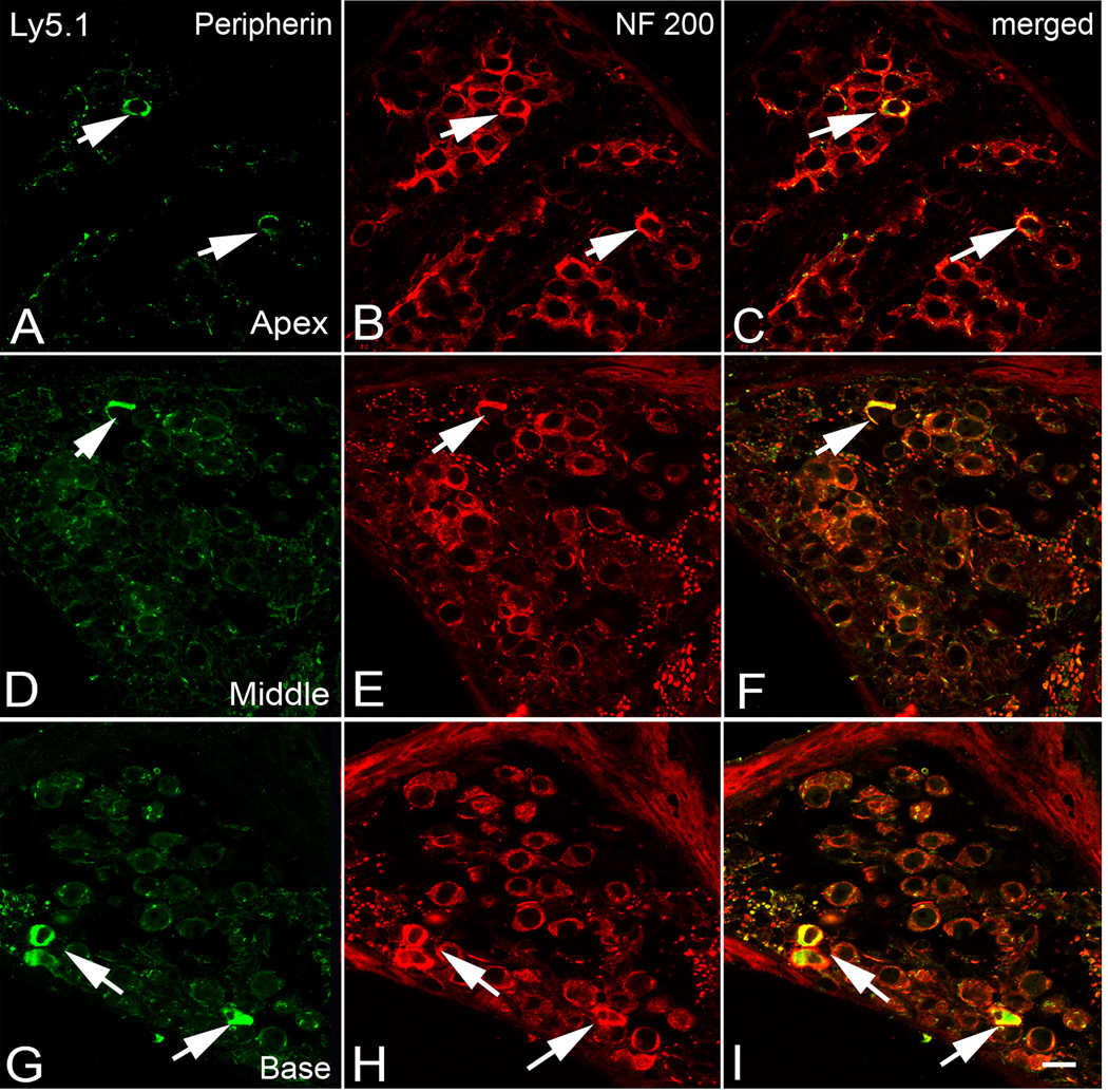
Figure 6. Dual immunolabeling for peripherin (green) and NF 200 (red) in the apical (A–C), middle (D–F) and basal turn (G–I) of an Ly5.1 mouse.
Neurons expressing both peripherin and NF 200 (arrows) are considered to be type II SGNs and are scattered along the periphery of Rosenthal’s canal. Scale bar: A–I, 10 µm. A magenta-green copy of this figure is available as Supplementary Figure 5.

