
Figure 6.
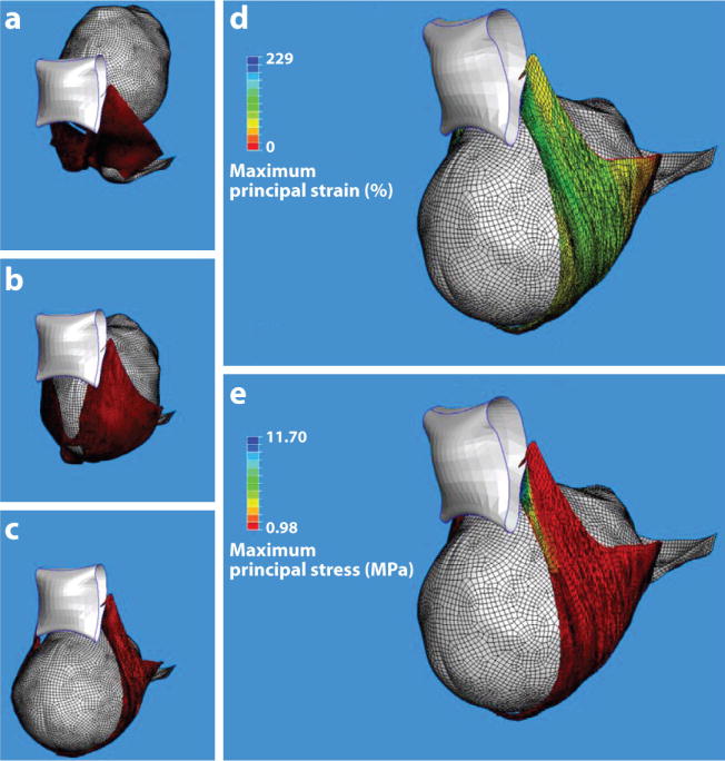
Finite element model results for a simulated single (120 N) push delivery for a 31-year-old mother and fetus at 40 weeks gestation. (a–c) An inferior three-quarter view of the pelvic floor viscohyperelastic soft tissue deformations at three stages during the second stage of labor: (a) Station +2, (b) a middle station, and (c) delivery of the fetal head. The levator ani muscle (red), fetal head (gray), and public bone (white) are shown. (d) The maximum predicted principal strain and (e) stress distributions in the pelvic floor soft tissues at time of delivery. The largest principal strain (d) reached 259% (3.59 stretch ratio). The blue region nearest the pubic bone indicates the local region of highest stress (e), corresponding to the location of muscle defects observed on magnetic resonance scans. Reproduced from Reference 12.