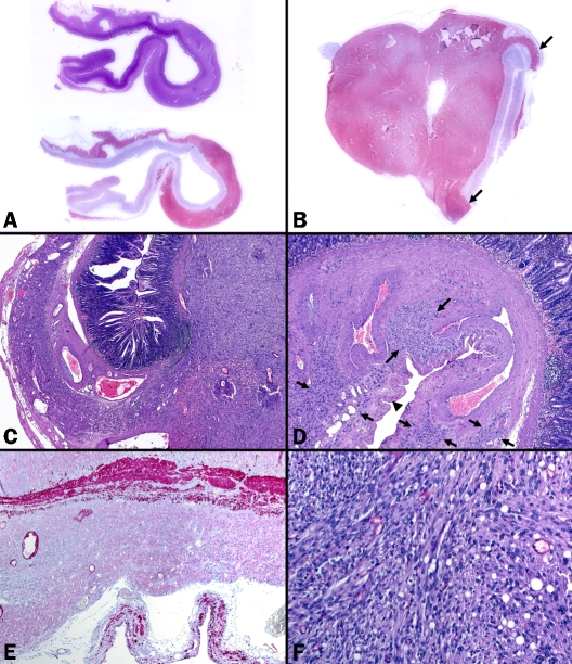
Figure 1.
Histological features of gastrointestinal stromal tumor in case 1. (A) Whole mount section from the diverticu-lar component (actual section length 2.5 cm) showed diffuse submucosal growth highlighted by CD117 (upper H&E, lower CD117). (B) Solid tumor component at the tip of a diffuse saccular structure (arrows) highlighted in the CD117 immunostaining. (C) Interphase between the two components, note dysplastic vessels. (D) Dysplastic vessels and intervening attenuated muscle fibers and connective tissue replaced the normal muscularis propria, note hypocellu-lar intervening tumorous tissue (between arrows). Prominent serosal undulations (arrow head) indicated thinning of the gut wall. (E) The muscularis propria was almost completely absent in the diffuse component (desmin stain). (F) Higher magnification of the solid tumor showed fascicular growth of spindled and rounded cells with occasional vacuoles.

