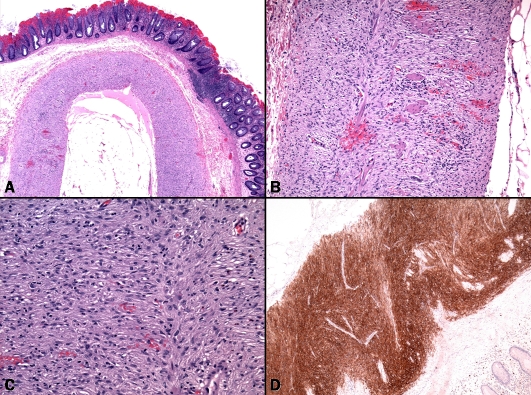
Figure 2.
Pathological findings in case 2. (A) Diffuse spindled proliferation replacing both layers of the muscularis propria in otherwise architecturally unremarkable colonic wall. (B) Residual muscle fibers of the muscularis propria were seen between tumor cells, note circumscribed growth towards both submucosa (left) and subserosa (right). (C) Tumor cells at higher magnification. (D) Diffuse staining with CD117 highlightingthe longitudinal pattern of tumor.

