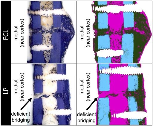
Fig. 5.
Typical histological appearance of healing in the far cortical locking (FCL) and locked plating (LP) groups, shown with toluidine blue stain and after image processing for callus differentiation (green = callus, blue = cortex, and pink = fibrous tissue). Three of six locked plating specimens had deficient bridging at the near cortex, while all other near and far cortices had bridged.

