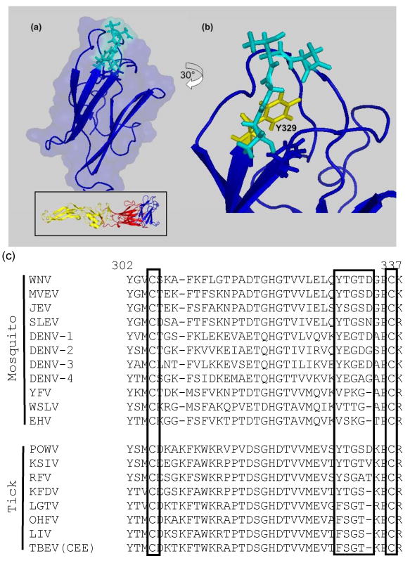
Figure 1.

Location of BC loop (residues 329-333, in cyan) in the WNV E domain III: (a) lateral view of domain III, oriented as for the complete E protein structure (inset); (b) rotated 30° to the left to show the location of Y329 (in yellow). (c) Amino acid sequence alignment of flavivirus domain III residues equivalent to 302-337 of WNV; flavivirus-conserved cysteines and BC loop residues 329-333 are boxed. Mammalian tick-borne flaviviruses are ordered according to their proximity to the root of that branch in previous phylogenetic analyses of their evolution (Gaunt et al., 2001; Grard et al., 2007) showing replacement of tyrosine with phenylalanine and shortening of the BC loop.
